
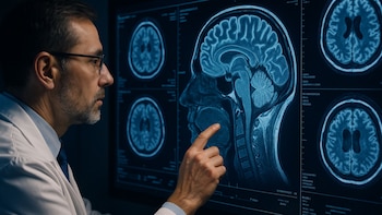
An international team of scientists has made a significant breakthrough in understanding how the human brain deals with fear. This research, recently published in Nature Human Behaviour, identified the essential coordination between deep regions of the human brain that facilitates the formation of new memory fingerprints during the process of extinguishing fear.
The scientists believe that this research opens the door for optimizing therapies aimed at treating anxiety disorders by revealing specific mechanisms that interrelate with behavioral adaptation and clinical treatment .
Conducted at the Pitié Salpêtrière hospital in Paris and the China South University of Guangzhou Hospital, this study involved intracranial recordings from 49 epilepsy patients. The collaboration spanned across Spain , Germany , France , and China , employing intracranial electroencephalography (IEEG) , which allows for high-resolution monitoring of activities in areas such as the amygdala , hippocampus , and prefrontal cortex . This advanced technique surpassed traditional non-invasive methods by offering detailed insight into the brain’s fear-learning pathways.

One of the study’s major revelations is that extinction of fear does not erase the original memory; instead, it fosters the creation of a new inhibitory memory that is heavily context-dependent. This new memory enables a coordinated response from the amygdala , hippocampus , and prefrontal cortex to suppress previously learned fear responses.
During the research, participants were shown images of objects believed to be electric, some of which were associated with an aversive stimulus —a loud cry paired with a negative facial expression. In the first phase, they learned to link certain images to threats. Later, during the extinction phase, these associations were modified, allowing researchers to observe how well patients could adjust their expectations and responses in light of the change in stimuli.
The findings indicate that all stimuli were presented in various visual contexts using an ABC paradigm to evaluate the contextual specificity of the memories . The behavioral results demonstrated that participants learned and updated their threat responses relative to the stimuli and contexts; however, complete elimination of fear was not achieved, emphasizing the persistence of the original memory while showing the coexistence of both memory traces .
From a neurophysiological perspective, the research team identified an increase in theta oscillations (4–12 Hz) in the amygdala during the extinction phase when considering stimuli viewed as safe. This finding was interpreted as a specific safety signal pertaining to the context of extinction, differing from earlier studies where increased theta was predominantly linked to fear acquisition.

The study also showed that neuronal representations of safe stimuli were more stable in the amygdala and temporal sensory regions during extinction , indicating effective memory consolidation contingent on specific contexts. Analysis of brain coordination revealed that synchronization between the amygdala, hippocampus, and lateral prefrontal cortex intensified during extinction, particularly when stimuli were presented in the learning context.
The lateral prefrontal cortex’s contextual specificity was correlated with a higher likelihood of reinstating fear memory later. According to the researchers, the interplay between fear memory and extinction memory holds significant clinical implications, influencing phenomena such as fear renewal and extinction recovery.
The importance of contextual specificity in extinction memory cannot be overstated. When extinction memory is context-dependent, fear can return in altered environments (“renewal”). Conversely, if the extinction memory becomes dominant, safety responses are more generalized.
The experts involved in the study stated, “The contextual specificity during extinction learning predicts the reappearance of fear memory footprints, while the reappearance of extinction memory footprints predicts safety responses.” This understanding opens the door for developing more effective treatments for anxiety and fear-related disorders, emphasizing the brain’s complex mechanisms involved in fear management .